
Category: Anatomy Models
Showing all 8 results
-
Buy via WhatsApp
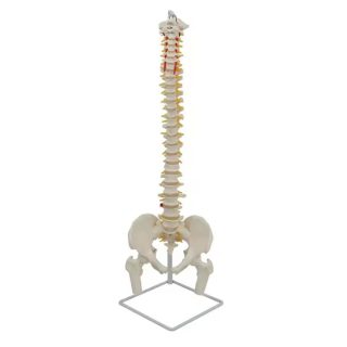
45cm Human Spine Model, Durable White
₨ 10,000Product description
Descriptions:
This spine model shows all the main features of each bone in greatdetail, including the spine, nerve roots, vertebral arteries, divided discs,transverse processes of the spine, and vertebral sections. It is an great teaching models for chiropractic, orthopedic, and other specialties.Features:
1. Made of synthetic materials that are durable and unbreakable.
2. Stable metal support frame with 5 rollers.
3. Close to the real weight of about 200 pieces of bones.
4. The limbs can be removed quickly and easily.
5. Natural bone size with 3 parts of assembled skull
-
Buy via WhatsApp
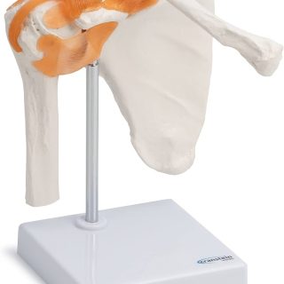
Anatomical model of the shoulder joint
₨ 7,500A life-size model of the shoulder joint, demonstrating its anatomy and biomechanics. It includes the scapula, clavicle, a portion of the humerus, and the joint ligaments. This highly flexible model clearly demonstrates abduction, anteversion, retroversion, and internal and external rotation.
-
Buy via WhatsApp
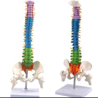
Color Human Spine Model 45cm Color Human Spine Model
₨ 10,000- SUITABLE FOR TEACHING—Our color human spine model is very suitable as a school teaching tool for learning and demonstration. It can also be used as a collection for decorating your laboratory and enriching your laboratory equipment.
- PREMIUM MATERIAL—Our color human spine model is made of high quality PVC material, with color, nonflammability, high strength, weather resistance and excellent geometric stability, easy to learn.
- CLEAR STRUCTURE—This high quality color human classic human spine model has medical details and a clear structure, allowing you to easily identify various parts of the spine, which is very suitable for teaching.
- DESCRIPTION DETAILS—A detailed description of all the main features of each spine, including the spine, nerve roots, vertebral artery, intervertebral disc, spine and transverse spine, femur, which can be used in medicine, exhibitions, art drawings, etc.
- UNUSED—We guarantee that the spine model is 100% brand new and quality guaranteed.
-
Buy via WhatsApp
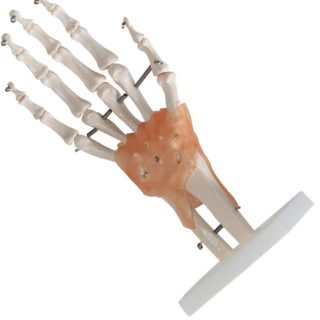
High Quality Pvc Human Skeleton Hand Joint Model
₨ 7,500-
Description:
- (Hand articulation model) – This model shows the anatomical structure of the hand and can show various functions of the hand. Contains flexible artificial ligaments. This model is suitable for use as intuitive pedagogical help for the medical interpretation of human anatomy.
- ➤ (Hardware) – This functional hip articulation model is made from a very sustainable non-toxic PVC material, very durable. Environmentally friendly materials, handmade, fine execution, excellent manufacture.
- ➤ (Easy to learn) – placed on the base, lightweight and portable. Helps students understand the morphology and construction of the hand articulation. Displays all the functions of the hand and external anatomical structures, allowing the patient or the student to better understand.
- ➤ (anatomically accurate) – The skeleton model of the human hand offers practitioners and students with incredible details and descriptive details of the hand and wrist structures, including a complete human hand and the lower parts of the cubitus and radius. The bones of the hand are maintained together with high quality wire that also allows flexibility and mobility.
- ➤ (variety of uses) – is suitable for anyone interested in anatomy, nursing, physiology. Ideal for children’s education. Applicable to schools, hospitals, physical health education. It will also be an excellent addition to your laboratory supplies.
-
-
Buy via WhatsApp
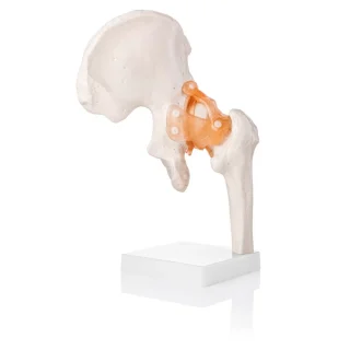
Hip Joint Model (Life-Size) With Ligaments
₨ 7,500Product Description
Hip Joint Model (Life-Size) With Ligaments – Anatomical Accuracy Meets Clinical Clarity
The hip joint is a marvel of stability and strength, bearing the weight of the body while allowing for fluid movement in multiple planes. The Life-Size Hip Joint Model with Ligaments brings this biomechanical wonder into focus with a clear, detailed view of bony and connective structures that define this critical joint.
Cast from an original human specimen, this model features the femoral head, acetabulum, ilium, ischium, and pubis—accurately aligned to show the true spatial relationship of the hip bones. What elevates this model further is the inclusion of ligaments such as the iliofemoral, pubofemoral, and ischiofemoral ligaments, presented in semi-flexible synthetic material to illustrate their role in joint stabilization and movement limitation.
This is not just a visual aid—it’s a functional teaching tool trusted by orthopedic educators, physiotherapists, medical students, and rehabilitation specialists. Mounted on a durable white base, it’s compact enough to sit on any clinic desk yet detailed enough to support high-level instruction and explanation.
One of the most common concerns in clinical education is showing patients or students how the hip’s deep structure contributes to pain, stiffness, or limited range of motion. This model answers that need. Whether it’s a labral tear, hip impingement, arthritis, or post-surgical explanation, this anatomical replica supports better understanding with every angle.
Search-optimized terms like “hip joint anatomy model,” “life-size pelvis and femur with ligaments,” “orthopedic hip replica,” and “functional ligamentous hip teaching aid” all match the purpose this model fulfills—precise, accessible, and clinically relevant education.
Made from high-quality, non-toxic PVC with durable ligament fibers, the model is lightweight, easy to clean, and built to last. If you’re aiming to clarify how the hip works—or why it sometimes doesn’t—this model is a quiet but powerful partner in every consultation or classroom.
-
Buy via WhatsApp
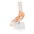
Human Foot Joint Model with Ligaments Scientific Life Size
₨ 7,500Human Foot Joint Model with Ligaments Scientific Life Size with Premium Display Base Best Teaching Tool for Patient Education & Anatomy StudyProduct Description- Skeletal Ankle model shows conditions such as ankle ligament strain, plantar fasciitis, achilles tendonitis, high ankle strain, metatarsal fracture and hallux dysfunction
- The practitioner or educator can show realistic movements with the flexible ligaments of the model.
- Also helps in educating patient and students about the normal and abnormal ankle, foot and toe joint ranges of motion as well as foot pronation and supination
- The foot skeleton shows a cross section of the bones including the periosteum and spongy bone
- The model is life size and clearly shows all the main anatomical structures of the ankle and foot
- The skeletal model is on base for better display and demonstration.
- The foot can be removed for close study and examination.
- Perfect for patient education, aspiring medical students, therapists, chiropractors, fitness trainers, etc. With Detailed Study Guide
-
Buy via WhatsApp
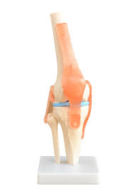
Knee Joint Model
Original price was: ₨ 8,500.₨ 7,500Current price is: ₨ 7,500.Product Description
Knee Joint Model:
Knee Joint Model with flexible and functional ligaments is great for in-depth study of human knee anatomy and demonstration purposes extremely useful for Doctors, Clinicians, Medical Students, Orthopaedics and Physiotherapists for demonstrating the functioning of human knee to their patients and students.
Knee Joint Model is one of the most popular anatomical model among physiotherapists, orthopaedics and chiropractors, It’s extremely useful for teaching, learning and patient education.
It is made with the precise focus on anatomical accuracy and robust functionality which allows our knee joint model to be used repeatedly without breaking any ligament.
Each anatomical structure from the cartilage to the ligament is shown accurately and precisely for accurate demonstration and studies.
Features:
- Knee Joint Model is of life size which helps in replicating real human knee structures for realistic depiction of human knee.
- Knee Joint Model features every tiniest anatomical detail which sets it apart from other available anatomy models in the market.
- Best anatomical model for teaching, self study, demonstration and patient education.
- Tibia, Femur and Patellar are present with the flexible ligaments which allows to mimic natural knee joint movement.
- Mounted on a sturdy base for ease of demonstration and learning.
-
Buy via WhatsApp
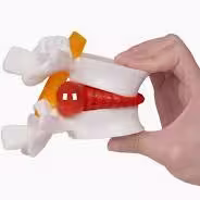
Lumbar Disc Herniation Anatomy
₨ 7,500Lumbar Vertebrae Model Anatomical Spine Lumbar Disc Herniation Anatomy Teaching Tool Lumbar Vertebrae Model
1. This model uses a fifth-segment lumbar vertebra and a fifth lumbar vertebrae, which are composed of a soft elastic body, which is convenient for the user to demonstrate, lumbar disc herniation, and shows the changes of the human intervertebral disc when the human body bends when it is distorted, 2. This model is widely used in orthopedics, surgery, for clinical teaching, occupational disease prevention, ergonomics, physical education, etc. It has good auxiliary teaching and demonstration 3. Made of high-quality PVC, and it is easy to wash. 4. Lumbar Vertebrae Model only, there accessories demo in the picture is not included. brand new and high quality Specifications: Type: Lumbar Vertebrae Model Color: As shown Material: PVC Package Contents: 1 * Lumbar Vertebrae Model Only the above package content, other products are not included. Note: Light and different displays may cause the color of the item in the picture a little different from the real thing. The measurement allowed error is +/- 1-3cm. Note: Due to the light and screen setting difference, the item’s color may be slightly different from the pictures. Please allow slight dimension difference due to different manual measurement.
Know Us
We deal in all kind of Physiotherapy, rehabilitation, surgical, acupuncture, hijama, and fitness equipments. Cash on delivery service all over the Pakistan.
Product Showcase